radius and ulna anterior and posterior view
Distally it articulates with the ulna again at the distal radioulnar jointIt forms part of the wrist joint by articulating with the scaphoid at its. Negative ulnar variance describes a state where the ulna is abnormally shortened compared to the radius and plays an important role in wrist pathology.
Ulna An Overview Sciencedirect Topics
A fall onto an outstretched hand is the.

. Cost-Utility Analysis of Thumb Carpometacarpal Resection Arthroplasty. Hand and Wrist Bones Quiz. Large protuberance on the proximal end of the ulna.
Rib. Maximum forearm pronation results in an increase in positive. There is a significant association between negative ulnar variance and Kienböck disease although the majority of people with negative ulnar variance do not have this conditionA causal association is difficult.
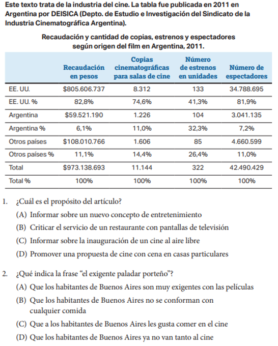
Os Coxa Bone Quiz Medial View Markings. Right clavicle superior view. Fractures of the radius and ulna are the most common fractures of the upper extremity with distal fractures occurring more often than proximal fractures.
Superficial anterior forearm muscles. Long slender bone with a hook at the proximal end that forms the elbow joint with the humerus. In this article we shall look at the anatomy of the anterior triangle of the neck its borders contents and subdivisions.
The superficial anterior forearm muscles share a common. Muscle contraction has to do with the bonding of two proteins called actin and myosin. The palm is maintained flat against the cassette.
Atlas C1 Axis C2 Cervical. Posteriorly and medially it is bounded by the limbus of the sphenoid bone. Os Coxa Bone Quiz Anterior Markings.
Figure 8-5a The Right Radius and Ulna Olecranon Proximal radioulnar joint ULNA Ulnar head Styloid process of ulna Posterior view Radial head Neck of radius RADIUS Ulnar notch of radius Styloid process of radius. It plays important role in wrist pathology such as ulnar impaction syndromes and thinning of the triangular fibrocartilage complex. View the Online Exams.
Right ulna and radius anterior and posterior views A. Radius and Ulna Bones Quiz. It is bounded as follows.
Explore myosin molecules and thick filaments actin molecules and thin filaments the organization of myosin. It is important to note that all triangles mentioned here are paired. The anterior cranial fossa consists of three bones.
Magnitude Direction Temporal Patterns and Frequency of Loss of Distal Radius Fracture Reduction in Women 50 years and Older. The superficial anterior forearm muscles are a group of five muscles located in the anterior flexor compartment of the forearmThese muscles include the pronator teres flexor carpi radialis flexor carpi ulnaris palmaris longus and flexor digitorum superficialis. Ulnae or ulnas is a long bone found in the forearm that stretches from the elbow to the smallest finger and when in anatomical position is found on the medial side of the forearm.
An interactive quiz covering anterior view of Hand and Wrist Bones through multiple-choice questions and featuring the iconic GBS illustrations. Scapula Posterior Aspect Scapula Lateral Aspect Humerus Proximal End Humerus Distal End Ulna. A Health Economic Study Using.
Femur Bone Quiz Anterior Markings. The routine minimal evaluation for distal radius fractures must include two views-a postero-anterior PA view and lateral view. The limbus is a bony ridge that forms the anterior border of the prechiasmatic sulcus a groove.
Patella Bone Quiz Anterior and Posterior Markings. The anterior triangle is a region located at the front of the neck. They are located on both the left and the right sides of the neck.
The frontal bone ethmoid bone and sphenoid bone. It runs parallel to the radius the other long bone in the forearmThe ulna is usually slightly longer than the radius but the radius is thicker. Proximally the head of the radius articulates with the capitulum of the humerus and the radial notch of the ulna at the elbowThe articulation between the radius and the ulna at the elbow is known as the proximal radioulnar joint.
The PA view should be obtained with the humerus abducted 90 degrees from the chest wall so that the elbow is at the same level as the shoulder and flexed 90 degrees. Anteriorly and laterally it is bounded by the inner surface of the frontal bone. Os Coxa Bone Quiz Lateral View Markings.
Femur Bone Quiz Posterior Markings. Positive ulnar variance describes where the distal articular surface of the ulna is more distal when compared to the articular surface of the radius. Therefore the radius is considered to be the larger.
Lies medially in the forearm when the body is in anatomical position.